Oral Surgeon
Uses CBCT imaging to evaluate bone volume, anatomy, nerve location, sinus position, surgical options, and implant placement considerations.
 Regional Implant Centers
Advanced 3D Imaging for Full-Mouth Rehabilitation
Mobile Cone Beam CT technology helps our specialists plan, coordinate, and deliver full-mouth rehabilitation with greater confidence and precision.
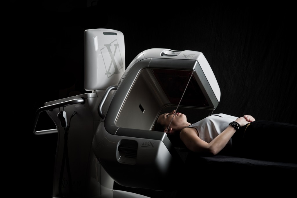
The better the planning, the better the outcome. At Regional Implant Centers, Mobile Cone Beam CT technology is used as part of a comprehensive planning and treatment workflow for patients considering full mouth dental implants, full arch dental implants, and complex full mouth rehabilitation.
Most dental offices use imaging primarily to diagnose a problem. Our team uses CBCT imaging to understand anatomy, support communication between specialists, guide dental implant planning, and help make more informed decisions before, during, and after treatment. The goal is not simply to capture images. The goal is to give the oral surgeon, prosthodontist, anesthesiologist, and laboratory technicians clearer information as they work together through the RISE Protocol™.
Simple Explanation
Cone Beam CT, often called CBCT imaging, is a form of 3D dental imaging that gives the clinical team a three-dimensional view of the mouth, jaw, and surrounding structures. Traditional dental x-rays can be helpful, but they usually show anatomy in a limited two-dimensional way. For routine dental needs, that may be enough. For full mouth dental implants and dental implant surgery, more information is often needed.
A Cone Beam CT scan can help the team evaluate bone structure, jaw anatomy, sinuses, nerves, existing teeth, and possible implant sites. This gives the doctors a more complete view of the areas involved in treatment. Patients do not need to understand every technical detail. What matters is that CBCT imaging helps the team see important anatomy more clearly before decisions are made.
Why It Matters
Full-mouth rehabilitation involves more than replacing teeth. The team must think about implant positioning, available bone, bite forces, facial support, restorative design, anesthesia needs, and the long-term function of implant-supported teeth. These decisions are connected. A surgical decision can affect the final smile. A restorative goal can influence where implants should be placed.
3D imaging for dental implants gives the team more information than traditional imaging alone. It helps the specialists evaluate where implants may be positioned, whether bone reconstruction may be needed, and how the surgical plan can support the final restoration. This is especially important for patients with failing teeth, missing teeth, previous dental work, dentures, infection, or significant bone loss.
Mobile Workflow
Traditional CBCT units are usually fixed in one imaging room. Mobile Cone Beam CT technology can be brought directly into treatment environments when appropriate. For a comprehensive implant center, that flexibility can support a smoother workflow and give the team timely access to imaging information when it is clinically useful.
The patient benefit is not about the machine itself. The benefit is how the technology supports efficiency, team communication, and treatment coordination. When imaging can be integrated into the clinical workflow, the specialists can review information together with fewer interruptions and a clearer shared understanding of the case.
During Treatment
One important advantage of Mobile Cone Beam CT technology is the ability to obtain imaging during treatment when clinically appropriate. In selected cases, this can help the team evaluate implant positioning, review anatomical relationships, and support additional decision-making while treatment is underway.
The ability to evaluate treatment during a procedure may help our team make more informed decisions. It can also improve communication between the oral surgeon, prosthodontist, and restorative planning team because everyone can review the same information in the context of the overall treatment goal. This does not replace clinical judgment, experience, or careful planning. It gives the specialists another layer of information when that information may be helpful.
Read full articleSupporting The RISE Protocol™
Technology supports collaboration, but it does not replace clinical expertise. The value comes from specialists using the same diagnostic data to plan treatment together.
Uses CBCT imaging to evaluate bone volume, anatomy, nerve location, sinus position, surgical options, and implant placement considerations.
Uses imaging and digital treatment planning to connect implant placement with the final bite, smile design, facial support, and implant-supported teeth.
Benefits from coordinated planning and case information when supporting patient comfort and safety during complex full-mouth rehabilitation.
Use improved planning information to support restorative design, provisional teeth, final prosthetic planning, fit, function, and esthetics.
Patient Benefits
Mobile Cone Beam CT helps patients understand that planning is part of treatment, not a separate step.
The Regional Implant Centers Difference
Dental implant technology alone does not create a successful outcome. The difference is how the technology is used. At Regional Implant Centers, Mobile Cone Beam CT is one component of a larger specialist-led workflow built around the RISE Protocol™.
Our approach brings imaging, digital treatment planning, oral surgery, prosthodontics, anesthesia support, and laboratory collaboration into one coordinated plan. That means the team is not simply reacting to problems as they appear. They are using better information to plan the path toward healthier function, a more confident smile, and a long-term rehabilitation strategy.
Schedule a CBCT-Guided ConsultationFrequently Asked Questions
Cone Beam CT is a type of 3D dental imaging that helps the team view bone structure, jaw anatomy, sinuses, nerves, teeth, and potential implant sites in three dimensions.
A regular dental x-ray usually provides a two-dimensional image. CBCT imaging provides a three-dimensional view, which can give the team more information for dental implant planning and full-mouth rehabilitation.
3D imaging for dental implants helps the team evaluate anatomy, bone availability, implant locations, and surgical considerations before treatment begins.
Cone Beam CT is recommended when the clinical team determines that 3D imaging is appropriate for diagnosis, planning, or treatment decisions. The need depends on each patient’s situation.
When clinically appropriate, Mobile Cone Beam CT may allow the team to obtain imaging during treatment to help evaluate implant positioning and support more informed decisions.
CBCT imaging gives the oral surgeon, prosthodontist, anesthesiologist, and laboratory team shared three-dimensional information during the Imaging and planning phases of the RISE Protocol™, helping full-mouth rehabilitation stay coordinated from diagnosis through surgery and restoration.