Advanced Implant Technology
Photogrammetry, Intraoral Scans and Guided Surgery: How Full-Arch Teeth Are Made to Fit, for Houston Patients
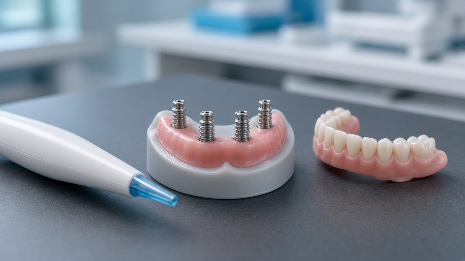
Photogrammetry, intraoral scanning and guided surgery do different jobs in full-arch treatment. Here is what each one means for how your new teeth fit.
Photogrammetry, intraoral scanning and guided surgery come up in almost every full-arch implant consultation in the Houston area, and they often get lumped together as "digital dentistry." They are not the same thing. Each one records or controls a different part of the job, at a different point in treatment, and only some of them have much to do with how well your final bridge fits. This article is for patients in Katy, Cypress, Sugar Land, Fulshear, Richmond and the rest of the Houston area who want to understand what these tools do before they sit down at our Katy office or anywhere else.
We have a separate article on guided implant surgery, which goes deep on surgical guides, accuracy research and the limits of guided placement. This one covers the step that comes after the implants are in: how the exact position of each implant is captured and handed to the lab, and why that record matters for a one-piece bridge.
This is general patient education. Your own records and steps depend on your anatomy, your implants and the plan your team recommends.
The short answer
Guided surgery helps put the implants where they were planned. Intraoral scanning records the shape of your gums, bite and teeth. Photogrammetry measures where each implant sits in relation to the others, which is the measurement a one-piece full-arch bridge depends on most. Research on photogrammetry for full arches looks favorable, but most of it comes from lab studies, and no single tool replaces the try-in visits and checks that confirm fit before the final teeth are made.
Three tools, three different jobs
These tools work at different stages and answer different questions, so asking which one is "best" usually misses the point.
| Tool | What it records or controls | When it is usually used | What it affects most |
|---|---|---|---|
| CBCT scan | 3D image of bone, nerves and sinuses | Before surgery, sometimes during | Where implants can safely go |
| Surgical guide (guided surgery) | Drill position, angle and depth | During implant placement | How closely surgery matches the plan |
| Intraoral scanner | Surface shape of gums, teeth and bite | Before surgery and after implants are placed | Tooth design, bite and how the bridge meets the gums |
| Photogrammetry | Position and angle of each implant relative to the others | After implants are placed | Whether a rigid bridge can seat without strain |
A simple way to think about it: the CBCT scan and the guide deal with where the implants go into the bone. The intraoral scan and photogrammetry deal with recording where they actually ended up, so the lab can build teeth that match. Even a well-guided surgery still needs an accurate record afterward, because the bridge has to fit the implants you have, not the ones in the plan.
What "fit" means for a full-arch bridge
A fixed full-arch bridge is one rigid piece screwed onto four or more implants. It should seat on all of them at once without being forced, which dentists call a passive fit.
Natural teeth move slightly in their sockets. Implants fused to bone do not. A single implant crown has room for a little error. A full-arch bridge spans the whole jaw, so a small error at one implant can mean the bridge rocks, or seats only when the screws pull it down. Dentists worry that this kind of strain can show up later as loose screws, chipped or cracked teeth, or stress on the implants.
How much misfit actually causes harm is less settled than marketing suggests. A 2017 systematic review by Katsoulis and colleagues found the evidence too limited to show that misfit damages bone around implants, but two clinical studies did point to a slightly higher risk of screw-related problems. The authors' conclusion was practical: the tolerable amount of misfit is still debated, and clinicians should aim for the least misfit possible. That is the job these recording tools and checkpoints are meant to do.
Intraoral scanning: the shape of your mouth
An intraoral scanner is a small handheld wand that takes thousands of images as it moves around your mouth and stitches them into a 3D model. It is very good at surfaces and less reliable when it has to measure across a whole toothless arch.
Before surgery, a scan can record your existing teeth, your bite or your current denture, which gives the team a starting point for designing new teeth. After the implants are placed, the scanner records the gum tissue and small connectors, called scan bodies, attached to the implants. For many patients the scan replaces some or all of the trays of impression putty that used to be the only option, which is easier on people who gag.
The limitation shows up on long, smooth stretches of gum with no teeth to use as landmarks. The scanner builds the model piece by piece, and small stitching errors can add up from one side of the jaw to the other. A 2026 meta-analysis in the Journal of Prosthodontics (Alfaraj and colleagues, 37 studies) found that, overall, digital and conventional impressions for full-arch implant bridges were about equally accurate. Intraoral scanners on their own did not differ from conventional impressions, conventional methods were slightly favored when there were more than four implants, and the authors described the evidence as varied and without a clear consensus. Results also depend on the scanner, the scan bodies and the experience of the person scanning.
What it feels like: the wand is about the size of a thick electric toothbrush handle. You keep your mouth open while it moves over your gums and implants for a few minutes. There is no taste and no setting time.
Photogrammetry: where the implants are in relation to each other
Photogrammetry uses a camera held outside the mouth to photograph coded markers attached to each implant, then calculates the position and angle of every implant relative to the others. It does not capture gums or teeth, so it is paired with an intraoral scan or another record.
Because the camera sees all the markers at once from outside the mouth, it does not have to stitch its way across the arch the way an intraoral scanner does. That is the main reason it is used for full arches.
The research so far points in its favor, with caveats:
- A 2025 systematic review and meta-analysis by Pozzi and colleagues compared photogrammetry and intraoral scanning for full-arch implant records in 13 studies. Photogrammetry was more accurate in 10 of them. In the lab studies the authors pooled, photogrammetry was significantly more accurate, and they reported that intraoral scanner angle errors could exceed the 1-degree level they associate with a passive fit, while photogrammetry's stayed below it.
- A 2023 study by the same group took three records from each of 11 patients receiving zirconia full-arch bridges. Compared with a conventional plaster record, the average position error was about 88 micrometers (thousandths of a millimeter) for photogrammetry and about 137 for the intraoral scanner, and the average angle error was 0.38 degree versus 0.79 degree.
- A 2026 meta-analysis in the Journal of Prosthetic Dentistry (Altalla and colleagues, 14 studies) also found better trueness and precision with photogrammetry.
Now the caveats. Most of these studies were done on models in a lab, the clinical studies are small, and the devices keep changing. Both Pozzi papers still recommend a rigid prototype try-in before the final screw-retained bridge is made. Better measurement lowers one source of error. It does not remove the need to check the fit in your mouth.
What it feels like: small markers are screwed onto your implants, and a clinician holds a camera a short distance from your face for a minute or two. Nothing goes deep into your mouth other than the markers.
Where guided surgery fits in
Guided surgery is about placing implants, not recording them. A 3D-printed guide, planned from your CBCT scan and the teeth designed for you, steers the drill to a planned position, angle and depth.
A 2018 meta-analysis by Tahmaseb and colleagues (20 clinical studies, 2,238 implants) found guided placement averaged about 1.2 millimeters of error at the entry point, 1.4 millimeters at the tip and 3.5 degrees of angle, with less accuracy in fully toothless jaws than in partly toothless ones. The authors called that clinically acceptable in most situations but advised keeping a safety margin of at least 2 millimeters from structures like nerves.
Those numbers are in millimeters. The fit numbers above are in thousandths of a millimeter. That difference explains why a guide cannot stand in for an accurate record afterward. A guide can help the implants land close to where the teeth were planned. The bridge still has to be built to the implants' actual positions. Our guided implant surgery article covers what guides can and cannot do, and how freehand and guided placement compare.
The FDA notes that dental CBCT scans are used for implant planning and encourages patients and dentists to avoid unnecessary radiation, so it is fair to ask why a scan is being taken and whether an earlier one can be used.
How the pieces usually connect in full-arch treatment
In a typical digital full-arch workflow, records taken before surgery are used to design the teeth first, surgery places the implants, and new records taken afterward are used to build the bridge.
The exact order varies by office and by case, but the general sequence looks like this:
- Records before surgery. A CBCT scan, an intraoral scan of your teeth or denture, photos of your face and smile, and bite records.
- Teeth-first planning. The team designs where the new teeth should sit for your lips, smile and bite, then plans implant positions to support them. This is often called restorative-driven planning.
- Surgery. The surgeon places the implants freehand, with a guide, or with a mix of both, depending on anatomy and judgment.
- Temporary teeth. Qualifying patients may leave with a fixed temporary bridge. It is not the final bridge.
- Healing. The implants fuse with the bone over a period of months, and your gums settle.
- Final records. The implants' positions are recorded again, with an intraoral scan, photogrammetry, a conventional impression, or a combination.
- Try-in and checks. A prototype of the final teeth is tried in, and the fit is checked, before the final material is made.
- Final bridge. The final prosthesis is delivered, adjusted and followed up.
Our older overview of digital planning before implant surgery introduces the planning tools in step 1. Our article on temporary vs final teeth explains steps 4 through 8 in more detail, and the surgery-day article covers what happens at step 3.
The checkpoints that catch problems before the final teeth
No impression method is perfect, so careful full-arch workflows build in checks. The two most common are a verification jig and a prototype try-in.
A verification jig is a rigid frame that connects the implants in your mouth. If it seats on every implant without rocking, the lab model or digital file is likely accurate. If it does not, the record is redone before anything final is made. A 2025 review in the Australian Dental Journal (Wong and colleagues) describes these jigs as a quality-control step that cannot guarantee a passive fit but can catch errors before the final bridge is fabricated.
A prototype try-in is a trial version of your new teeth, often printed or milled in a plastic material. It lets you and your team judge tooth position, smile line, lip support, speech and bite in your own mouth while changes are still easy to make. Your dentist may also check how it seats with one screw tightened at a time.
If you are asked to come back for an extra try-in visit, that is usually a sign the team is checking its work, not a sign that something went wrong.
What technology does not decide
Precise records help, but how a full-arch bridge does over the years also depends on your bite, grinding habits, cleaning, health, materials, follow-up care and the people doing the work.
A bridge that fits well can still chip if you clench at night, collect plaque if it is hard to clean underneath, or loosen if screws are not checked. Our articles on teeth grinding with full-arch implants, cleaning and maintenance, loose or broken bridges and zirconia vs acrylic teeth cover those parts of the picture. Planning tools also do not make treatment right for everyone. Some people are better served by a snap-on overdenture or a well-made traditional denture.
How our team uses these tools at the Katy office
Our Houston-area office is at 707 S Fry Rd, Suite 150, in Katy, and our in-house dental lab is there. Our team plans full-arch cases with CBCT imaging, intraoral and facial scanning, photogrammetry and digital treatment planning.
Under the RISE Protocol, our prosthodontist plans the final teeth before surgery begins, and the surgical plan is built around them. Our SegMark Workflow brings CBCT imaging, intraoral scans, bite information and facial references into one digital plan, which supports clearer surgical guides and plans when guided planning is appropriate for your case. Whether a guide is used, and what kind, is a surgical decision made case by case.
Our photogrammetry page describes it as a way to capture the three-dimensional position of implants after placement so the restorative and lab teams can design a full-arch prosthesis aimed at a passive fit. It is typically used after implants are placed, and photogrammetry alone does not guarantee a result. Our mobile cone beam CT lets the team study bone, nerves and sinuses before surgery and capture updated scans during the procedure when the clinical team decides that is appropriate.
The in-house lab at Katy means the technicians who shape your teeth work beside your doctors and can see you in person, which helps with tooth shape, shade, lip support and smile line. Dr. Polyxeni Konti, our prosthodontist, serves patients at the Katy office.
Both of our offices offer traditional dentures, snap-on implant overdentures and fixed full-arch bridges, and zygomatic and pterygoid implants are performed by Dr. Dan Holtzclaw and Dr. Enrique Tabarini for patients with severe upper bone loss. Our full-mouth dental implants in Katy page and Katy location page cover the office itself, and our article for Sugar Land, Fulshear and Cypress patients covers getting there.
Treatment is not right for everyone, and a consultation is where the team decides what your case actually needs.
Questions to ask any Houston office about fit
- How will you record my implant positions for the final bridge: intraoral scan, photogrammetry, a conventional impression, or a combination?
- Will there be a prototype try-in, a verification jig, or both, before the final teeth are made?
- Who designs and makes the bridge, and will that person see me?
- Is a surgical guide planned for my case, and if not, why not?
- If the final bridge does not seat well, what happens next?
- How many visits should I expect between surgery and the final teeth?
- What maintenance visits and screw checks do you recommend after the bridge is delivered?
How insurance and financing fit in
Planning technology does not change how insurance works. At our office, medical insurance may help only with surgical professional services, and implants, the prosthesis and temporary teeth are billed to you.
Medical insurance may help only with surgical professional services, such as extractions, bone grafting and alveoloplasty. If you have a PPO plan with oral surgery coverage, it is worth checking whether it may help with those surgical parts of your care. Please verify your benefits with us and your carrier before you decide. Only PPO plans work for our out-of-network oral surgery benefits, and we cannot bill Federal Employee Program (FEP) Blue Cross Blue Shield plans. Our Insurance Advantage program includes a medical and dental benefit evaluation, and our insurance disclaimer explains why no estimate is a guarantee.
Before treatment, you receive a written plan with estimated insurance benefits, financing options and your expected out-of-pocket investment. Financing may be available for qualified patients through third-party financing partners, which set their own approval requirements and terms. After your final teeth, a Maintenance Package is required, with enrollment within 30 days of the final prosthesis, and our implant warranty page explains why it is not a lifetime guarantee.
Frequently asked questions
Is photogrammetry better than an intraoral scan for full-arch implants?
For recording where several implants sit relative to each other, recent reviews found photogrammetry more accurate in most studies, though much of the research was done in labs. It does not record gums or teeth, so it is usually paired with an intraoral scan rather than replacing it.
What does photogrammetry feel like for the patient?
Small markers are screwed onto your implants and a camera held outside your mouth photographs them for a minute or two. There is no impression putty and no wand moving across the back of your mouth for that step.
Is guided surgery the same thing as photogrammetry?
No. Guided surgery uses a 3D-printed guide to steer implant placement during surgery. Photogrammetry measures where the implants ended up afterward so the lab can build the bridge to match. They solve different problems and can be used in the same case.
Will I still need a conventional impression?
Possibly. Some teams use intraoral scans and photogrammetry alone, some combine them with a conventional impression or a verification jig, and some choose based on the case. Ask how your final records will be taken and checked.
Does digital technology mean my bridge will not need adjustments?
No. Precise records reduce one source of error, but bite adjustments, a prototype try-in and follow-up visits are still normal. Long-term results also depend on your bite, cleaning, health and maintenance.
Talk with our Katy team about planning your teeth
If you are comparing full-arch options in the Houston area and want to know how your new teeth would be planned and checked for fit, bring your questions to a consultation. Qualified patients receive a complimentary consultation, an advanced CBCT scan and an insurance benefits evaluation. Schedule a consultation by phone or in person at our Katy office, or call (832) 632-7037.
Sources & Resources
- Pozzi A, Arcuri L, Carosi P, et al. Photogrammetry Versus Intraoral Scanning in Complete-Arch Digital Implant Impression: A Systematic Review and Meta-Analysis. Clinical Implant Dentistry and Related Research, 2025.
- Pozzi A, Carosi P, Gallucci GO, et al. Accuracy of complete-arch digital implant impression with intraoral optical scanning and stereophotogrammetry: An in vivo prospective comparative study. Clinical Oral Implants Research, 2023.
- Altalla H, Alhelou H, Karaduman F, et al. Comparative accuracy of photogrammetry and intraoral scanners in recordings for complete arch implant-supported prostheses: A systematic review and meta-analysis. Journal of Prosthetic Dentistry, 2026.
- Alfaraj A, Alqudaihi F, Khurshid Z, et al. Comparative analyses of accuracy between digital and conventional impressions for complete-arch implant-supported fixed dental prostheses: A systematic review and meta-analysis. Journal of Prosthodontics, 2026.
- Katsoulis J, Takeichi T, Sol Gaviria A, et al. Misfit of implant prostheses and its impact on clinical outcomes. Definition, assessment and a systematic review of the literature. European Journal of Oral Implantology, 2017.
- Tahmaseb A, Wu V, Wismeijer D, et al. The accuracy of static computer-aided implant surgery: A systematic review and meta-analysis. Clinical Oral Implants Research, 2018.
- Wong AWH, Nedelcu R, Hamilton A. Clinical Recommendations for Implant Verification Jigs in Analogue and Digital Workflows. A Narrative Review. Australian Dental Journal, 2025.
- U.S. Food and Drug Administration. Dental Cone-beam Computed Tomography.
This article is general patient education and is not medical or dental advice, a diagnosis or a treatment plan. Research on photogrammetry, intraoral scanning and guided surgery is still developing, much of it comes from laboratory studies, and results vary by device, operator and case. No planning or recording technology guarantees fit, comfort or long-term success. Same-day teeth are temporary and offered only to qualifying patients. Implant treatment is not appropriate for every patient, outcomes vary, and no surgery or anesthesia is free of risk. Insurance coverage is never guaranteed, and financing is provided by independent third-party companies. Regional Implant Centers has offices in Katy and Hutto, Texas.
Ready to Learn What Is Possible?
Schedule a consultation with Regional Implant Centers.
Schedule ConsultationFrequently Asked Questions
Is photogrammetry better than an intraoral scan for full-arch implants?
For recording where several implants sit relative to each other, recent reviews found photogrammetry more accurate in most studies, though much of the research was done in labs. It does not record gums or teeth, so it is usually paired with an intraoral scan rather than replacing it.
What does photogrammetry feel like for the patient?
Small markers are screwed onto your implants and a camera held outside your mouth photographs them for a minute or two. There is no impression putty and no wand moving across the back of your mouth for that step.
Is guided surgery the same thing as photogrammetry?
No. Guided surgery uses a 3D-printed guide to steer implant placement during surgery. Photogrammetry measures where the implants ended up afterward so the lab can build the bridge to match. They solve different problems and can be used in the same case.
Will I still need a conventional impression?
Possibly. Some teams use intraoral scans and photogrammetry alone, some combine them with a conventional impression or a verification jig, and some choose based on the case. Ask how your final records will be taken and checked.
Does digital technology mean my bridge will not need adjustments?
No. Precise records reduce one source of error, but bite adjustments, a prototype try-in and follow-up visits are still normal. Long-term results also depend on your bite, cleaning, health and maintenance.
This content is educational and not a substitute for a consultation.